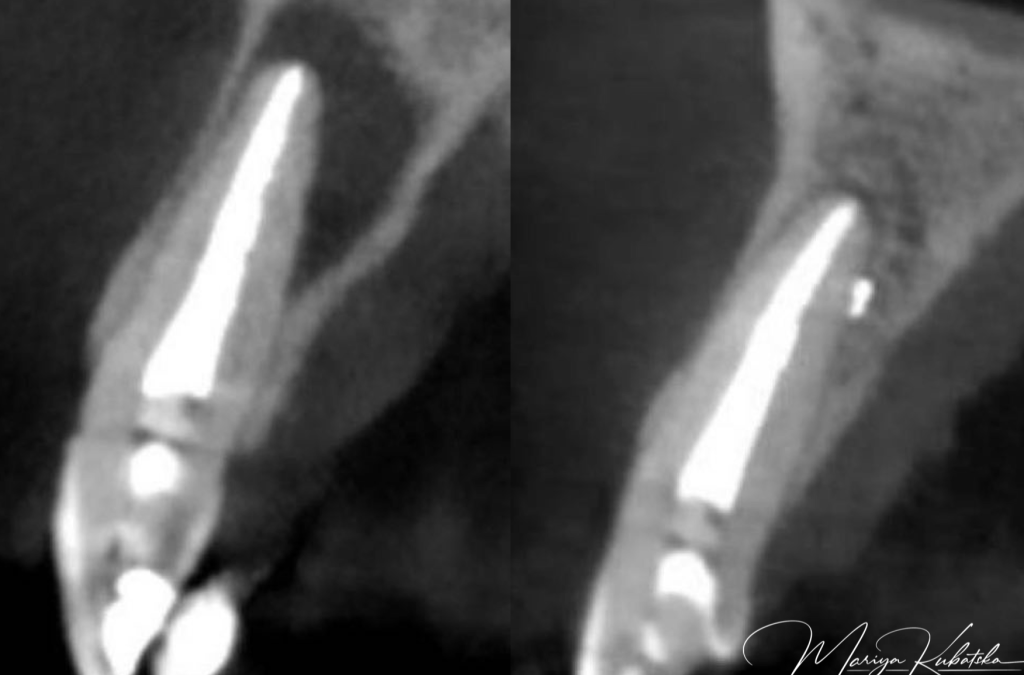
Apical periodontitis serves an essential protective function aimed at confining bacteria discharged from the root canal space and preventing them from spreading into adjacent bone marrow spaces and other remote sites. The process is unique in the sense that it cannot eradicate the source of infection. The reason is that once a pulp has become necrotic, defence mechanisms cannot operate far into the root canal owing to the lack of vascular support. Although these mechanisms can act at the apical margins of the necrotic tissue, they are unable to penetrate it in a fully developed tooth. Consequently, without proper endodontic treatment, apical periodontitis may prevail chronically. Bone resorption is a most conspicuous feature of apical periodontitis and is an unavoidable side-effect of the defensive process. Some may view it as a “price” paid by the host to provide the necessary, effective immune response to the root canal infection. On a microscopic level, different structural frameworks of apical periodontitis can be identified. These forms include apical granuloma, apical abscess, and apical cyst. Clinically and radiographically, these histopathological entities cannot be distinguished from each other or recognised, with the exception of abscesses with a sinus tract. Apical granuloma is the most common form of apical periodontitis and consists of an inflammatory lesion dominated by lymphocytes, macrophages, and plasma cells. Apical abscess denotes the presence of pus within the lesion. Abscess formation may reflect a shift in cellular dynamics within a pre-existing apical granuloma or be a direct outcome of an acute primary infection. An apical cyst is an epithelium-lined cavity that contains fluid or semi-solid material and is commonly surrounded by dense connective tissue variably infiltrated by mononuclear leukocytes and PMNs.Apical cysts are divided into pocket cysts (bay cysts) and true cysts. A pocket cyst is an apical inflammatory cyst that contains a cavity that is open to and continuous with the root canal space. True apical cysts, on the other hand, are located within the periapical granuloma with no apparent connection between their cavity and that of the root canal space.root and mesial region of the distal root of the mandibular molars. The cause of forming C-shaped canals and the prevalence of the danger zone in C-shaped roots is a failure of Hertwig’s epithelial root sheath to fuse on the lingual or buccal root surface. If Hertwig’s epithelial root sheath fails to fuse on the lingual surface, then the groove and danger zone occur on the lingual side of the root. If the root sheath fails to unite on the buccal surface-danger zone appears on the buccal side.
Click here to read the rest of my article posted in endodontics.styleitaliano.org

